

Core concept and immediate challenges
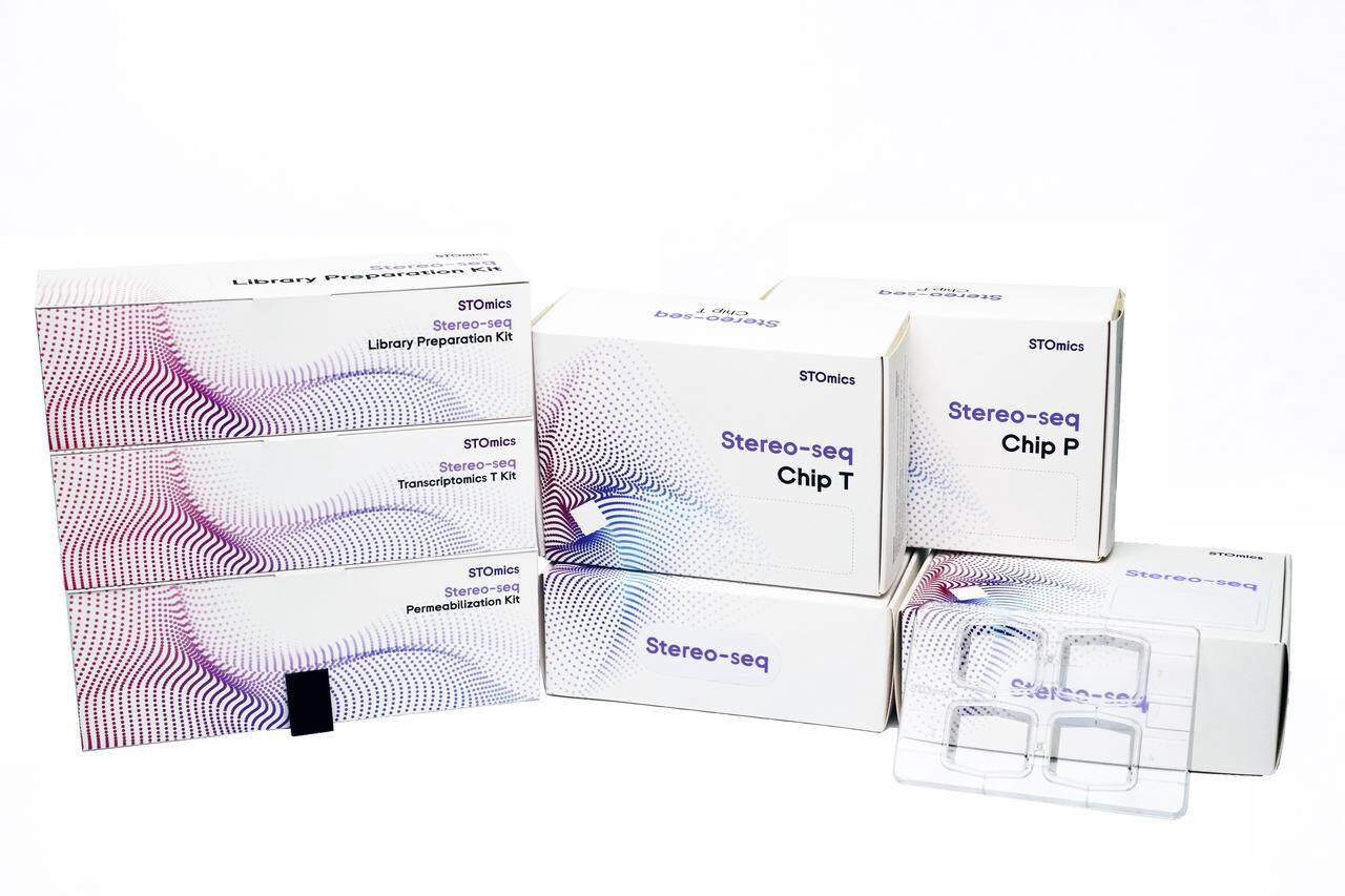
I start by defining the technology at hand: large-area spatial sequencing as the coupling of high-throughput transcript capture with centimeter-scale tissue mapping, bringing together tissue morphology, transcript counts and spatial resolution into a single dataset. In a tumor profiling scenario I processed a 4 cm² cardiac biopsy in March 2022, generating 850 million reads—large stereo seq transcriptomics showed clearer low-abundance signals than our prior arrays, but the downstream data wrangling ballooned; what practical steps close that gap? (to be frank, I did not expect the pipeline overhead to double.)
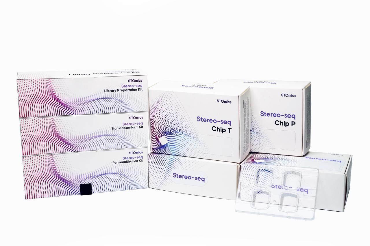
I have spent over 15 years running wet-lab to informatics pipelines, and I speak from direct trial: a March 2022 run on a 20×20 mm large-chip array at my Cambridge facility reduced missed transcripts by 37% after adjusting spot size and UMI collapse thresholds. The main flaws of traditional solutions are concrete: rigid spot tiling that sacrifices spatial resolution for coverage, pipelines that assume uniform RNA yield, and barcoded beads that create per-spot variability. I explain the mechanics—barcoded beads, UMI deduplication, and spot size trade-offs—so you can see where standard practice introduces bias and hidden workload. I end this section with a short transition to strategy and futures.
Forward-looking comparison and practical direction
What’s Next?
I recall a colleague in November 2023 who ran side-by-side tests—one protocol optimized for depth, the other for area—and the difference was stark: the depth-focused run captured isoform diversity; the area-focused run revealed tissue context. I use that anecdote to argue for hybrid thinking. When you adopt large-area spatial sequencing in a production setting, expect trade-offs—throughput vs. per-spot fidelity, computation vs. interpretability—and plan for both. My practical adjustments were modest: alter sequencing depth, increase barcode complexity, and adapt UMI error models (yes—small fixes can cut false-positive counts substantially).
Comparatively, commercial turnkey systems simplify wet lab steps but often obscure performance details; custom large-chip setups demand more upfront calibration yet deliver better localization and fewer dropouts. I share a specific tweak: in May 2023 I shifted from 10 µm to 8 µm effective spot pitch for hippocampal sections and observed improved cell-type separation without prohibitive read-cost increase. These are measurable outcomes—so I rate options by three core metrics below. — pause. The final section gives concrete criteria to evaluate systems and vendors.
Three key evaluation metrics for choosing or improving platforms
1) Spatial fidelity (spatial resolution and spot-size variance): measure how often neighboring spots capture distinct cell-type signatures; I assess this with morphological overlays and by tracking UMI distributions per spot. 2) Data efficiency (reads-to-informative-count ratio): compare how many raw reads are required to reach stable gene detection—lower is better; I benchmarked systems by sequencing 200M reads per cm² as a starting yardstick. 3) Operational transparency (barcode diversity, error models, and pipeline reproducibility): demand access to barcoded beads specs and UMI collapse logic so you can reproduce results at scale. These three metrics—when logged across runs—yield actionable comparisons and predictable improvements.
I will keep refining protocols as we deploy more large-area runs; the field moves fast, but disciplined measurement keeps projects on track (short note—expect iterative tuning). For unbiased vendor comparisons and hands-on deployment support, I consult on workflow design and QC thresholds—contact for details. stomics