
Starting point: the problem that keeps folks up at night
Everyone’s got a hang-up: will the scan capture the margin, will the lab read my bite right, will the crown seat first try? I’ve been around enough chairs and bench tops to know those worries sound the same from Mobile to Memphis. I even saw shining 3d dental units set up on demo tables at the Greater New York Dental Meeting, and teams were swapping notes about a digital dental solution that tightened handoffs. After years of helping clinics and labs streamline cases, here’s a problem-driven look at the questions that matter and practical fixes that actually work.
Problem: scan accuracy and margin capture
What goes wrong: poor soft-tissue retraction, shiny restorations confusing the scanner, and missed margins. Fix it: use retraction cord or a quick hemostatic, dry and angle the scanner deliberately, and take a focused close-up sweep of the margin before backing off. If the restoration’s shiny, a light dusting or matte spray for the lab model photo cuts through the glare. Don’t assume one pass is enough — a targeted rescan beats guessing later.
Problem: file formats and interoperability
What goes wrong: lab says it can’t open your file, or margins shift after export. Fix it: export an uncompressed STL or PLY depending on the lab’s preference, include a labeled bite file, and keep a copy of the original scanner project file. Talk to your lab up front: ask which file they prefer and request a short test case. Open systems save headaches; closed systems can be fine if everyone’s agreed on the export routine.
Problem: occlusion and implant analogs
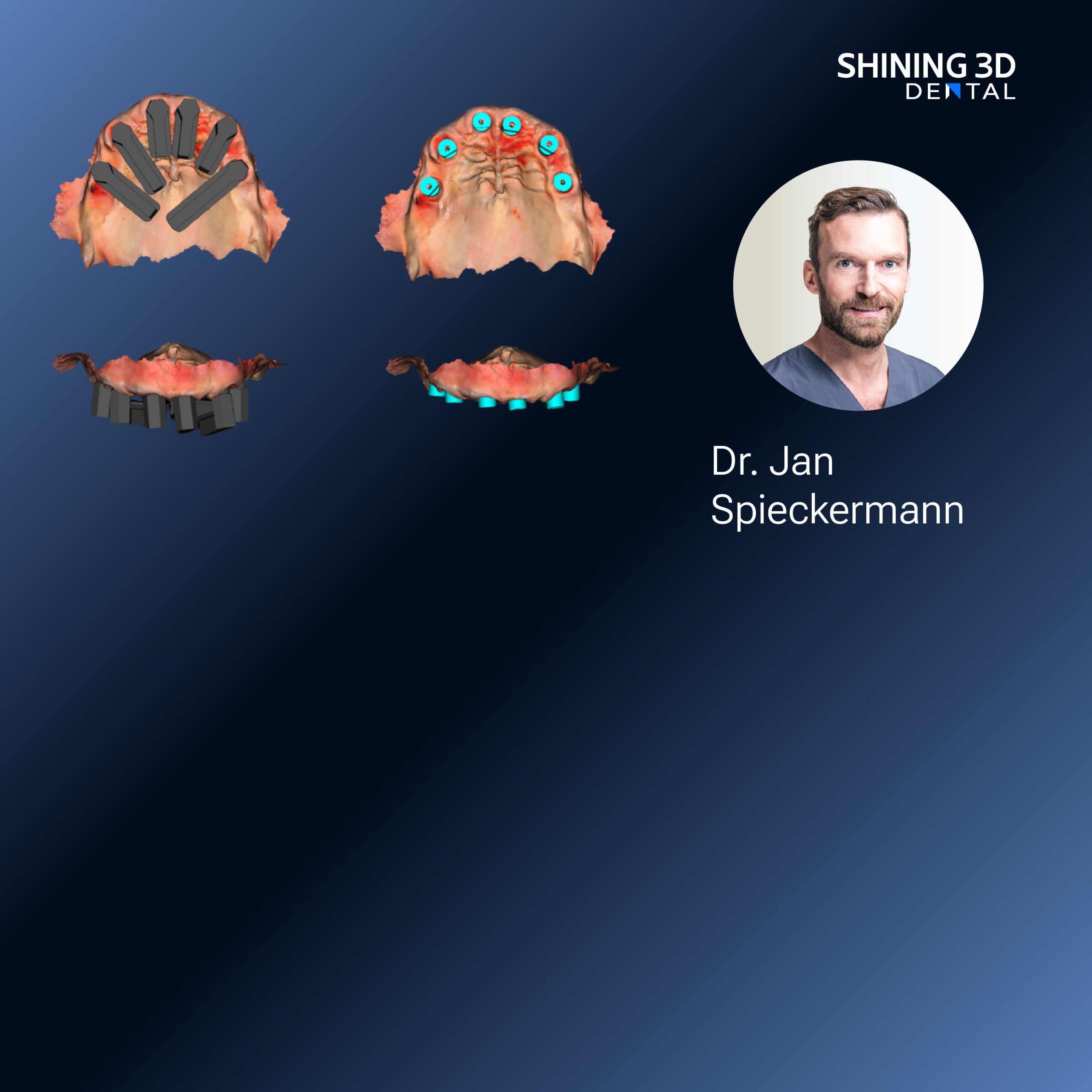
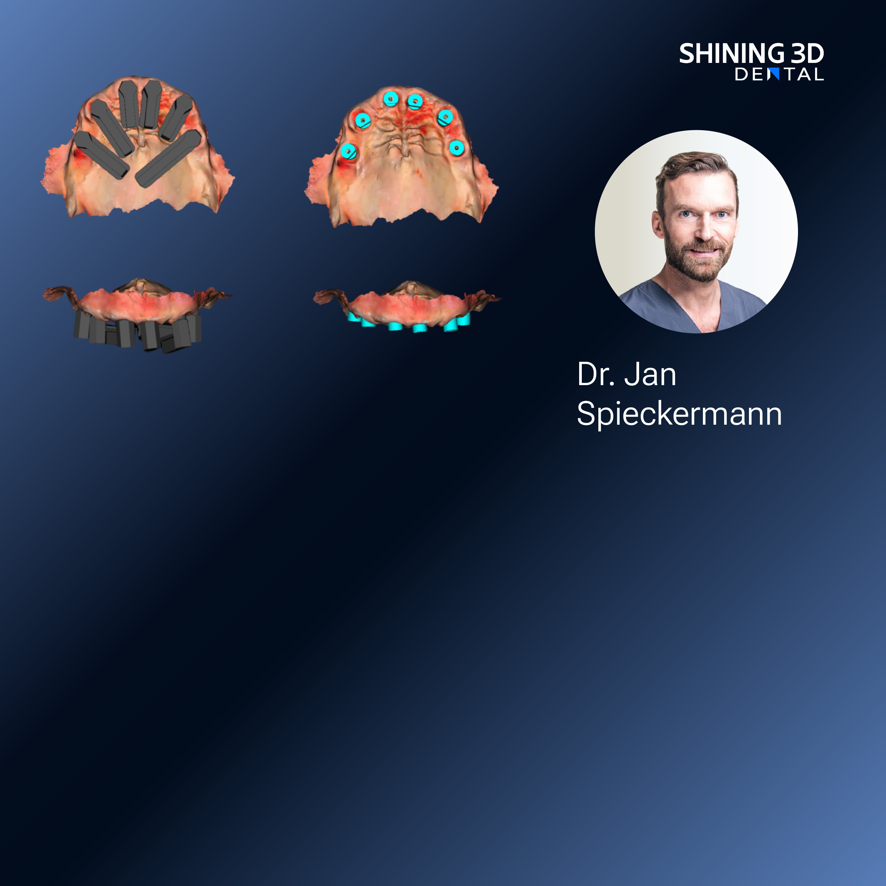
What goes wrong: crowns look perfect solo but don’t fit the bite. Fix it: capture a proper bite registration with teeth seated firmly together, then verify occlusion in multiple scans — canine, molar, and a light centric stop. For implant cases, send scan bodies that match the lab’s library and include clear photos of hex orientation. A labeled diagram saves phone calls and remakes.
Choosing gear and software — real trade-offs
What to weigh: speed versus accuracy, cost versus service, closed versus open software. Intraoral scanners differ more in workflow than in basic accuracy; pick one your team actually wants to use every day. Labs want consistent files and library compatibility. Consider service agreements and updates — a cheap scanner that sits broken won’t save money. If you’re comparing options, look at ease of integrating with your lab and whether the software handles library updates cleanly.
Handoff habits that cut remakes
What goes wrong: missing case notes, unlabeled models, or poor shade communication. Fix it: standardize a checklist — patient name, arch side, material request, margin photos, and shade tabs. Send a quick voice note or photo if something’s unusual. Labs appreciate a single, well-labeled ZIP over five scattered emails. Set minimum image requirements and filename conventions so nothing gets lost between systems.
Common mistakes labs and clinics keep making
Thinking hardware alone will fix workflow. Relying on default settings. Skipping simple verification steps. The cure: run a calibration check monthly, create a short staff SOP for scanning angles and bite captures, and do a weekly review of remakes to spot patterns. Train consistently — a well-trained assistant will save way more time than the fanciest gadget.
Wrap-up: what helps the most
Problems get smaller when teams use clear steps and agree on formats. Be deliberate with margin capture, standardize your exports, and label everything so no one’s left guessing. When clinics and labs want reliable gear that fits those habits, many turn the practical parts of their workflow toward trusted systems like SHINING 3D DENTAL because they match the routines that actually keep cases moving and patients smiling.